

Demetrios G. Vavvas, Nancy Huynh,
Louis Pasquale and Eliot L. Berson.
Massachusetts Eye and Ear Infirmary.
Department of Ophthalmology
Harvard Medical School | Boston, Massachusetts, USA
Acta Ophthalmol. 2010: 88: 156–157
2008 The Authors Journal compilation
ª 2008 Acta Ophthalmol
doi: 10.1111/j.1755-3768.2008.01359.x
A70-year-old woman presented with a complaint of diminishing vision over 2 years, which was worse in the left eye (OS). She had received hydroxychloroquine treatment for discoid lupus for approximately 15 years at doses of < 400 mg⁄ day (< 6.5 mg⁄ kg ⁄ day; cumulative dose of ≈ 2.2 kg).
The medication was discontinued in 2000 at the onset of ocular symptoms. Serial visual fields (VFs) (Fig. 1) revealed progressive loss that had prompted one ophthalmologist to initiate treatment for normal-tension glaucoma.
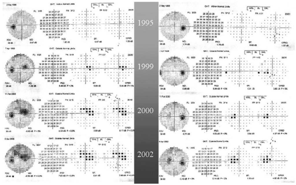
Fig. 1. Humphrey visual field (VF) findings in a 70-year-old woman with hydroxychloroquine retinal toxicity showing progressive VF loss from 1999–2002. The patient stopped hydroxychloroquine in 2000.
Fluorescein angiography (FA) in 2000 (Fig. 2A) showed areas of hyperfluorescence in the parafoveal region, leading to an alternative diagnosis of juxtafoveal telangiectasia.
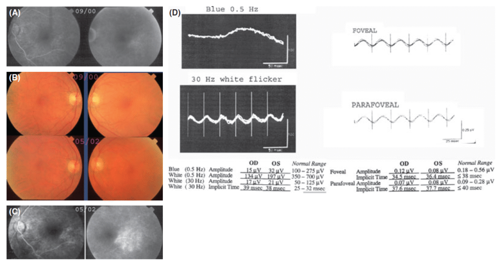
Fig. 2. (A) Fluorescein angiogram (FA) in 2000 (OS) with early and late frames showing hyperfluorescence in the parafoveal area. (B) Fundus
On examination, the patient’s visual acuity was 20 ⁄ 20 OD and 20 ⁄ 60 OS. No afferent pupillary defect was seen. Intraocular pressures
were 14 mmHg OD and 15 mmHg OS. No colour deficit on Ishihara plates was found. Examinations of anterior segments, optic discs and
maculae were normal (Fig. 2B). Repeat VF testing and FA findings were similar to those obtained previously (Fig. 2A, C). Scotopic and
photopic electroretinography (ERG) revealed suppressed and delayed photoreceptor responses, suggestive of widespread outer retinal disease that is unexpected in glaucoma (Fig. 2D). Hydroxychloroquine is an antimalarial drug frequently used in the treatment of haematological diseases.
One of its potentially adverse sideeffects is irreversible maculopathy, although this is rare relative to the number of patients on the drug (Levy et al. 1997). Early signs of hydroxychloroquine toxicity can present as a paracentral scotoma detected by threshold VF testing. Advanced toxicity is typically characterized by bull’s eye maculopathy associated with retinal pigment epithelium (RPE) atrophy (Easterbrook 1999).
Our patient developed hydroxychloroquine toxicity over 2 years after discontinuation of the medication, with incomplete bull’s eye
lesions only apparent on FA. The pattern of VF loss mimicked an arcuate defect seen in glaucoma. However, it did not respect the horizontal meridian despite adequate patient fixation, as shown by the position of the blind spot (Fig. 1).
Finally, glaucoma, as a disease that involves the ganglion cells, does not affect full-field or focal ERG amplitude responses to single flashes of light. Our patient’s ERG was diminished, which is in accordance with hydroxychloroquine toxicity (Weiner et al. 1991).
The FA findings, showing RPE changes and the absence of telangiectatic vessels or leakage, were also consistent with the diagnosis of hydroxychloroquine retinopathy, but not with juxtafoveal telangiectasis.
References
Easterbrook M (1999): Detection and prevention of maculopathy associated with antimalarial agents.
Int Ophthalmol Clin 39: 49–57.
Levy GD, Munz SJ, Paschal J, Cohen HB, Pince KJ & Peterson T (1997): Incidence of hydroxychloroquine retinopathy in 1207 patients in a large multicentre outpatient practice. Arthritis Rheum 40: 1482–1486.
Weiner A, Sandberg MA, Gaudio AR, Kini MM & Berson EL (1991): Hydroxychloroquine retinopathy. Am J Ophthalmol 112: 528–534.