
Electric Car Radiation

Electric Car Radiation

Electric Car Radiation: What You Need to Know

Electric cars are changing the way we move, offering cleaner mobility and reduced reliance on fossil fuels. But many drivers and passengers wonder: do electric cars emit harmful radiation?

What kind of radiation do electric cars produce?

Electric vehicles (EVs) rely on high-voltage batteries, inverters, and motors.

These systems generate low-frequency electromagnetic fields (ELF-EMFs) whenever the car is in motion. In addition, features such as Bluetooth, Wi-Fi, GPS, and mobile connectivity create radiofrequency (RF) signals the same type of radiation your smartphone or home Wi-Fi router uses.

Are these radiation levels dangerous?

According to the World Health Organization (WHO) and the International Commission on Non-Ionizing Radiation Protection (ICNIRP), the radiation levels measured inside electric cars are well below international safety limits.


Research shows:

  • Exposure levels are often comparable to household appliances like induction cookers, laptops, or hair dryers.

  • The radiation is non-ionizing, meaning it does not damage DNA the way ionizing radiation (like X-rays) can.

  • No confirmed long-term health risks have been linked to EMF levels from electric cars.


Drivers vs. passengers: is there a difference?
  • Drivers sit closer to batteries and motors, so their exposure is slightly higher but still within safe ranges.

  • Passengers typically experience even lower exposure, since distance reduces EMF intensity.

Key takeaway

Yes, electric cars emit electromagnetic radiation, but at very low levels.

Current evidence shows it is safe for both drivers and passengers. The main considerations for EV ownership remain battery life, charging infrastructure, and overall safety not radiation.  Electric cars offer a safe, sustainable alternative to combustion engines. Radiation exists, but it is no greater risk than the technology we already use daily in our homes and offices.

This article is for informational purposes only and does not constitute medical or technical advice. Always refer to official guidelines from the WHO, ICNIRP, and your vehicle manufacturer for detailed safety information.

Radiation at Home: What You Really Need to Know

home radiation

Radiation at Home: What You Really Need to Know

The word radiation often triggers fear, but not all radiation is harmful. In fact, we live surrounded by different kinds of electromagnetic fields every day —from mobile phones and Wi-Fi routers to Bluetooth earbuds and microwave ovens. Understanding when radiation is actually harmful and when it is harmless  helps us use our devices with confidence rather than worry.

Ionizing vs. Non-Ionizing Radiation

Science makes a clear distinction:

  • Ionizing radiation: (like X-rays or nuclear radiation) carries enough energy to damage DNA and potentially cause health problems.
  • Non-ionizing radiation (like Wi-Fi, Bluetooth, mobile phones, microwaves, infrared heat) does not have that ability. Its main effect is mild heating, and at normal household levels it is considered safe.

In everyday life, we are almost exclusively exposed to non-ionizing radiation.

The Role of Distance

Radiation intensity follows the “ inverse square law ”: if you double the distance from the source, exposure drops to one-quarter.

At just a few meters away, most household devices emit levels that are negligible.

Device by Device

Mobile phones
The strongest source at home. When held against the ear, they can emit up to 1–2 watts. Using hands-free or speaker mode instantly reduces exposure by over 100 times.

Bluetooth earbuds
Despite concerns, they operate at just 1–10 milliwatts hundreds of times weaker than a phone call. Even with both earbuds in use, total exposure remains far below international safety limits.

Wi-Fi routers
Typically emit around 100 – 200 milliwatts. At a distance of a few meters, the exposure is minimal. Signals from neighboring apartments are thousands of times weaker.

Cordless phones (DECT)
The base station emits continuously, even when not in use. If placed on a bedside table, this can mean constant exposure. The simple fix: keep the base away from the bedroom or choose an Eco Mode model that switches off when idle.

Microwave ovens
Inside, they generate high power (500 – 1000 watts) to heat food, but the metal shielding prevents leaks. Regulations allow only tiny leakage, far below safety thresholds. Unless the door is damaged, they are safe to use.

Heaters and convectors
They emit only infrared heat radiation — the same type of harmless warmth our own bodies emit. No link to harmful radiation.

Hidden sources
Baby monitors, smart home devices, induction cooktops, and electric blankets also emit non-ionizing radiation. While generally safe, they can be sources of continuous exposure if placed right next to the body (e.g., a baby monitor by a crib).

Key Takeaways
  • Household devices emit non-ionizing radiation, which is safe under international limits.
  • The biggest exposure comes from a mobile phone at the ear.
  • Distance is your best shield: even small separation dramatically lowers exposure.
  • Sensible habits hands-free calls, keeping routers and cordless bases out of the bedroom, and ensuring microwaves are in good condition are enough to minimize any risk.

Bottom line: Radiation is part of modern life, but with a little awareness, we can keep our exposure well within safe levels and enjoy our technology without fear.

Πόσο μας βλάπτει η ακτινοβολία των συσκευών;

Radiation at Home: What You Really Need to Know

Ακτινοβολία στο σπίτι: Τι ισχύει για κινητό, Wi-Fi, Bluetooth, μικροκύματα και άλλες συσκευές

Η ακτινοβολία δεν θεωρείται επιβαρυντική για την υγεία του ανθρώπου όταν βρίσκεται κάτω από τα όρια ασφαλείας που έχουν οριστεί από διεθνείς οργανισμούς (π.χ. ICNIRP, Παγκόσμιος Οργανισμός Υγείας).

Συγκεκριμένα:

  • Χαμηλή ένταση (ισχύς εκπομπής)

Όταν η ενέργεια που απορροφά το σώμα είναι πολύ μικρή για να προκαλέσει βλάβη. Αυτό μετριέται ως SAR (Specific Absorption Rate).
Για κινητά τηλέφωνα, το όριο στην ΕΕ είναι 2 W/kg.

  • Μη ιοντίζουσα ακτινοβολία

Οι ακτινοβολίες που δεν έχουν αρκετή ενέργεια για να“σπάσουν” χημικούς δεσμούς στο DNA (όπως κάνει η ιοντίζουσα ακτινοβολία: ακτίνες Χ, γ, ραδιενέργεια).

  • Χρόνος έκθεσης

Ακόμα και χαμηλή ακτινοβολία μπορεί να θεωρηθεί προβληματική αν η έκθεση είναι συνεχής και σε υψηλές εντάσεις.

Όταν η χρήση είναι μέτρια και διαλείπουσα, ο κίνδυνος είναι πρακτικά μηδαμινός.

  • Απόσταση από την πηγή

Η ένταση πέφτει πολύ γρήγορα όσο απομακρυνόμαστε από την πηγή (κανόνας αντιστρόφων τετραγώνων).

Άρα, μια ακτινοβολία δεν επιβαρύνει την υγεία όταν:

  • Είναι μη ιοντίζουσα.
  • Η ισχύς/έκθεση είναι κάτω από τα διεθνή όρια ασφαλείας.
  • Ο χρόνος χρήσης είναι λογικός και δεν υπάρχει υπερβολική, συνεχής έκθεση.

Wi-Fi & Bluetooth

Το Bluetooth είναι μια τεχνολογία ασύρματης επικοινωνίας μικρής εμβέλειας που επιτρέπει σε συσκευές να συνδέονται μεταξύ τους και να ανταλλάσσουν δεδομένα χωρίς καλώδια.

Βασικά χαρακτηριστικά:

  • Εμβέλεια: Συνήθως 10 μέτρα (υπάρχουν και εκδόσεις με μεγαλύτερη εμβέλεια, μέχρι και 100 μέτρα).

  • Συχνότητα λειτουργίας: 2,4 GHz (ίδια μπάντα με Wi-Fi αλλά με διαφορετική τεχνολογία).

  • Κατανάλωση ενέργειας: Σχετικά χαμηλή, γι’ αυτό χρησιμοποιείται σε κινητά, ακουστικά, wearables κ.λπ.

  • Ταχύτητα μεταφοράς: Από μερικές εκατοντάδες kbps στις πρώτες εκδόσεις έως αρκετά Mbps στις πιο νέες.

Το Bluetooth και το Wi-Fi ανήκουν στη μη ιοντίζουσα ακτινοβολία, άρα δεν «σπάνε» το DNA και θεωρούνται ασφαλή όταν η ένταση είναι κάτω από τα διεθνή όρια. Η επιβάρυνση εξαρτάται από την ισχύ, τον χρόνο χρήσης και κυρίως την απόσταση· όσο αυξάνεται η απόσταση από τη συσκευή, η ακτινοβολία μειώνεται εκθετικά και γίνεται πρακτικά αμελητέα.

Bluetooth ακουστικά

  • Πολύ χαμηλή ισχύς

Εκπέμπουν σε επίπεδα της τάξης των 1–10 milliwatt (mW), δηλαδή εκατοντάδες φορές πιο αδύναμα από ένα κινητό που μιλάς στο αυτί (που φτάνει μέχρι 1–2 Watt).

  • Μη ιοντίζουσα ακτινοβολία

Όπως το Wi-Fi, δεν έχει ενέργεια αρκετή να βλάψει το DNA.

Η επίδραση είναι θερμική (παραγόμενη θερμότητα), αλλά σε τόσο χαμηλά επίπεδα που είναι πρακτικά αμελητέα.

  •  Χρόνος χρήσης

Η συνεχής, πολύωρη χρήση δεν έχει αποδειχθεί ότι βλάπτει, όμως για προληπτικούς λόγους προτείνεται εναλλαγή (να μη φοράς ακουστικά όλη μέρα χωρίς διακοπή).

  • Απόσταση

Το κινητό όταν μιλάς το κρατάς κολλητά στο κεφάλι, ενώ με τα Bluetooth ακουστικά το κινητό μένει στην τσέπη ή στο τραπέζι → αυτό από μόνο του μειώνει την έκθεση.

Συμπέρασμα:
Τα Bluetooth ακουστικά εκπέμπουν πολύ χαμηλότερη ακτινοβολία από τα κινητά και θεωρούνται ασφαλή για καθημερινή χρήση μέσα στα διεθνή όρια.

Ray-Ban Meta AI

Είναι «έξυπνα» γυαλιά με Bluetooth & Wi-Fi, μικρές κάμερες, μικρόφωνα, ηχεία κ.λπ.  Περνούν δεδομένα και επικοινωνούν ασύρματα με το τηλέφωνο ή το δίκτυο, δηλαδή εκπέμπουν ραδιοσυχνότητες (RF), όπως κάθε συσκευή που χρησιμοποιεί Bluetooth / Wi-Fi.

Πιθανοί κίνδυνοι / ακτινοβολία

Η ηλεκτρομαγνητική ακτινοβολία που εκπέμπουν είναι μη ιοντίζουσα, όπως για Bluetooth / Wi-Fi γενικά — δεν έχει ικανότητα να «σπάσει» χημικούς δεσμούς DNA. Υπάρχει μελέτη που δείχνει ότι η θέση της κεραίας στη συσκευή μπορεί να επηρεάσει το πόση ενέργεια απορροφάται από διαφορετικούς ιστούς στο κεφάλι.

Όσο μεγαλώνει η απόσταση από την πηγή (η κεραία στους βραχίονες των γυαλιών) τόσο μειώνεται η έκθεση.

Είναι ασφαλή;

Μέχρι σήμερα δεν υπάρχει αποδεδειγμένο ιατρικό στοιχείο ότι τα Ray-Ban Meta προκαλούν βλάβες όταν χρησιμοποιούνται κανονικά, κάτω από τα πρότυπα ασφαλείας για RF / SAR.

Wi-Fi router

Ένα τυπικό Wi-Fi router εκπέμπει 100–200 mW (0,1–0,2 Watt).
  • Αυτό είναι 10–20 φορές λιγότερο από ένα κινητό σε κλήση στο αυτί (1–2 Watt).
  • Η ισχύς πέφτει απότομα με την απόσταση (κανόνας αντιστρόφων τετραγώνων).
  • Στα 30 εκ. απόσταση: ~1/100 της ισχύος.
  • Στο 1 μέτρο: ~1/1000.
  • Στα 3 μέτρα: πρακτικά αμελητέο.
Ακτινοβολία από γειτονικά σπίτια
  • Τα σήματα Wi-Fi που “πιάνουμε” από τους γείτονες είναι συνήθως χιλιάδες φορές πιο αδύναμα από το δικό μας router. Γι’ αυτό και τα “πιάνουμε” με λίγες μπάρες σήμα → πολύ μικρή ένταση. Η συμβολή τους είναι σχεδόν αμελητέα σε σχέση με το Wi-Fi που έχουμε μέσα στο ίδιο μας το σπίτι.
Συγκριτικά
  • Κινητό στο αυτί σε κλήση: μέχρι 1–2 Watt → η μεγαλύτερη έκθεση.
  • Δικό μας router στο σαλόνι: ~0,1 Watt, αλλά πέφτει πολύ γρήγορα με την απόσταση.
  • Γειτονικά router: 1000 φορές πιο αδύναμα → πρακτικά ασήμαντη έκθεση.

Η κύρια πηγή ακτινοβολίας στο σπίτι είναι το δικό μας κινητό όταν μιλάμε στο αυτί, όχι το Wi-Fi router και σίγουρα όχι τα γειτονικά router.

Κινητό τηλέφωνο

  • Εκπέμπει όταν μιλάς ή όταν ψάχνει σήμα.
  • Ισχύς εκπομπής: μέχρι 1–2 Watt (1000–2000 mW).
  • Μετριέται σε SAR (Specific Absorption Rate). Στην ΕΕ το όριο είναι 2 W/kg.
  • Όταν είναι στο αυτί, έχεις τη μέγιστη έκθεση.
  • Όταν είναι 30–50 εκ. μακριά (π.χ. στο τραπέζι με hands-free), η ακτινοβολία πέφτει 100+ φορές.

Ασύρματο τηλέφωνο

  • Έχει βάση που είναι συνεχώς συνδεδεμένη με το ρεύμα και εκπέμπει συνεχώς, ακόμη κι όταν δεν μιλάς.

  • Ισχύς εκπομπής: ~10–250 mW ανάλογα με το μοντέλο.

  • Το ακουστικό επικοινωνεί με τη βάση με παρόμοια τεχνολογία με το κινητό, αλλά σε μικρότερη απόσταση.

  •  Αν η βάση είναι δίπλα στο κρεβάτι, έχεις συνεχή έκθεση όλο το 24ωρο (ακόμα κι αν δεν μιλάς).

  • Η βάση εκπέμπει συνεχώς για να κρατά “σύνδεση” με το ακουστικό, ακόμα κι αν δεν μιλάς.
  • Το ίδιο το ακουστικό, όταν είναι παρκαρισμένο πάνω στη βάση και φορτίζει, συνήθως δεν εκπέμπει ή εκπέμπει ελάχιστα (εξαρτάται από το μοντέλο).
  • Άρα η μόνιμη πηγή ακτινοβολίας είναι η βάση, όχι το ακουστικό όταν είναι κουμπωμένο.

Τι σημαίνει αυτό στην πράξη:

  • Αν η βάση είναι στο σαλόνι ή στο γραφείο, η έκθεση είναι μικρή γιατί η απόσταση βοηθά.
  • Αν η βάση είναι στο κομοδίνο δίπλα στο κεφάλι, έχεις συνεχή έκθεση όλη νύχτα (σαν να έχεις ένα μικρό Wi-Fi router δίπλα σου).

Κάποια νεότερα μοντέλα έχουν λειτουργία Eco Mode, όπου η βάση δεν εκπέμπει όταν το τηλέφωνο δεν χρησιμοποιείται.

Συμπέρασμα:

Όταν το ασύρματο είναι στη βάση του, η βάση εξακολουθεί να εκπέμπει συνέχεια, το ακουστικό όμως όχι. Η τοποθέτηση της βάσης (πόσο κοντά μας είναι) είναι ο πιο σημαντικός παράγοντας.

Φούρνοι μικροκυμάτων

  • Εκπέμπουν μικροκύματα γύρω στα 2,45 GHz (ίδια μπάντα με Wi-Fi, αλλά με πολύ μεγαλύτερη ισχύ).

  • Η ισχύς μέσα στο φούρνο είναι 500–1000 Watt για να θερμαίνει το φαγητό.


Τι γίνεται με την ακτινοβολία προς τα έξω
  • Ο φούρνος έχει μεταλλικό περίβλημα και ειδικό “δίχτυ” στο τζάμι που κρατά την ακτινοβολία μέσα.
  • Η διεθνής νομοθεσία επιτρέπει μόνο πολύ μικρή διαρροή: < 5 mW/cm² σε απόσταση 5 cm από την πόρτα.

Αυτό είναι χιλιάδες φορές μικρότερο από την ενέργεια που έχει μέσα στον φούρνο.

Όταν είναι σε καλή κατάσταση (χωρίς σπασίματα, χωρίς φθαρμένη πόρτα ή λαστιχάκια), η ακτινοβολία που “φεύγει” είναι αμελητέα και κάτω από τα όρια ασφαλείας.

Η απόσταση παίζει τεράστιο ρόλο:
  • Στα 30–50 εκ. από τον φούρνο η διαρροή είναι πρακτικά μηδενική.
  • Το φαγητό δεν μένει “ραδιενεργό”· απλώς θερμαίνεται επειδή τα μόρια νερού πάλλονται.
Συμπέρασμα

Ένας φούρνος μικροκυμάτων που λειτουργεί σωστά δεν επιβαρύνει την υγεία. Αν η πόρτα έχει σπάσει ή δεν κλείνει καλά, τότε μπορεί να υπάρξει διαρροή και χρειάζεται άμεση αντικατάσταση.

Θερμοπομποί (ηλεκτρικές θερμάστρες τύπου panel)

Πώς δουλεύουν οι θερμοπομποί

Είναι ηλεκτρικές συσκευές θέρμανσης που μετατρέπουν το ρεύμα σε θερμότητα.

Ζεσταίνουν τον αέρα μέσω αντίστασης και κυκλοφορίας (convection).

Δεν εκπέμπουν ραδιοκύματα ή μικροκύματα, αλλά μόνο θερμική ακτινοβολία (υπέρυθρη).

  • Τι ακτινοβολία εκπέμπουν

Η ακτινοβολία τους είναι υπέρυθρη θερμότητα — η ίδια που βγάζει μια σόμπα χαλαζία, μια λάμπα θέρμανσης ή ακόμα και το σώμα μας. Είναι μη ιοντίζουσα ακτινοβολία, άρα δεν βλάπτει DNA και δεν έχει σχέση με Wi-Fi ή κινητά. Η μόνη “επίδραση” είναι η αύξηση θερμοκρασίας γύρω από τον χρήστη.

  • Ασφάλεια για την υγεία

Όταν χρησιμοποιούνται σωστά, δεν υπάρχει κίνδυνος από ακτινοβολία. Το μόνο που χρειάζεται προσοχή:

  • να μην καλύπτονται με ρούχα (πυρασφάλεια),
  • να υπάρχει σωστός αερισμός στον χώρο,
  • να τηρούνται οι οδηγίες χρήσης.
Συμπέρασμα:


Οι θερμοπομποί δεν εκπέμπουν επιβλαβή ακτινοβολία. Εκπέμπουν μόνο υπέρυθρη θερμότητα, όπως κάθε θερμαντική πηγή. Ο μοναδικός πραγματικός κίνδυνος είναι πρακτικός (πυρκαγιά αν χρησιμοποιηθούν λάθος), όχι από την ακτινοβολία τους.

Οι πιο επιβλαβείς επισκευές.

  • Baby monitors (ασύρματα μόνιτορ μωρών)

Εκπέμπουν συνεχώς 24/7, συχνά με ισχύ συγκρίσιμη με router. Αν είναι δίπλα στο κρεβατάκι, το μωρό δέχεται σταθερή ακτινοβολία. Λύση: τοποθέτηση σε απόσταση.

  • Smart home συσκευές (Κάμερες, έξυπνοι λαμπτήρες, ασύρματα κουδούνια, συναγερμοί)

    Συνδέονται με Wi-Fi ή Zigbee/Z-Wave και εκπέμπουν μόνιμα χαμηλή ισχύ. Αν υπάρχουν πολλές, η “συνολική” έκθεση αυξάνει (αν και παραμένει πολύ χαμηλή σε σχέση με κινητό).

  • Ηλεκτρικές κουβέρτες / θερμαινόμενα στρώματα

    Παράγουν χαμηλής συχνότητας ηλεκτρικά και μαγνητικά πεδία όσο είναι στην πρίζα. Αν χρησιμοποιούνται για ώρες κολλητά στο σώμα, η έκθεση είναι συνεχής (όχι αποδεδειγμένα επικίνδυνη, αλλά πολλοί ειδικοί συστήνουν μέτρο).

  • Παλαιά ασύρματα τηλέφωνα (χωρίς Eco Mode)
    Μπορεί να εκπέμπουν συνεχώς και με περισσότερη ισχύ από τα νεότερα μοντέλα.

Anti-VEGF in Retinopathy of Prematurity, Need to Titrate

Identification of Nore1 as a Potential Ras Effector

Anti-VEGF in Retinopathy of Prematurity, Need to Titrate

Demetrios G. Vavvas
Angiogenesis Laboratory, Retina Service, Department of Ophthalmology, Massachusetts Eye and Ear Infirmary, and Massachusetts
General Hospital, Harvard Medical School, Boston, Massachusetts; Vavvas@meei.harvard.edu

Retinopathy of prematurity (ROP) is a major cause of blindness in children in the Western World. Premature birth disturbs normal vascular development. Children with more avascular retina have higher risk of developing pathologic neovascularization (NV) and
severe ROP. The present standard of care has been laser or cryoablation of the peripheral avascular retina in order to control the
pathologic neovascularization. Despite the undeniable benefit of these primitive yet time tested interventions, there is incomplete effectiveness and significant morbidity. Molecular understanding of normal and abnormal vascular development has highlighted the
role of VEGF and suggested a role of anti-VEGF therapy.1 The effectiveness of anti-VEGF therapy in AMD and other neovascular diseases has generated excitement about application of ‘‘gentler’’ anti-VEGF therapies for ROP. The Bevacizumab Eliminates the
Angiogenic Threat of Retinopathy of Prematurity study gave us excitement, but also caution about delayed normal vascular development and NV formation.(2)

It is natural to think that in a developing organ, the exact level of VEGF inhibition will be important in order to control pathologic NV, while at the same time allowing normal vascular development to proceed. Lutty et al.3 has demonstrated this principle in a dog model of ROP using an agent that blocks simultaneously VEGF-A, VEGF-B, and placental growth factor. In this issue, McCloskey et al.4 show similar findings using VEGF blockade alone. In a well controlled study in rodents, they show that more anti-VEGF blockade is not better but worse, leading to increased avascular retina and delayed atypical NV formation with activation of compensatory angiogenic signals that may not respond to subsequent VEGF blockade. They also showed that anti- VEGF therapy was associated with reduced weight gain. Their study highlights the need for more systematic research regarding the appropriate dose and monitoring of anti-VEGF in ROP babies in order to have a safer and more effective therapy than the current primitive ablative approaches.

References

1. Smith LE. Through the eyes of a child: understanding retinopathy through ROP the Friedenwald lecture. Invest Ophthalmol Vis Sci.
2008;49:5177–5182.
2. Mintz-Hittner HA, Kennedy KA, Chuang AZ, BEAT-ROP Cooperative Group. Efficacy of intravitreal bevacizumab for stage 3þretinopathy
of prematurity. N Engl J Med. 2011;364:603–615.
3. Lutty GA, McLeod DS, Bhutto I,Wiegand SJ. Effect of VEGF trap on normal retinal vascular development and oxygen-induced retinopathy in the dog. Invest Ophthalmol Vis Sci. 2011;52:4039–4047.
4. McCloskey M,Wang H, Jiang Y, SmithGW, Strange J, Hartnett ME. Anti-VEGF antibody leads to later atypical intravitreous neovascularization and activation of angiogenic pathways in a rat model of ROP. Invest Ophthalmol Vis Sci. 2013;54:2020–2026.

Identification of Nore1 as a Potential Ras Effector

Identification of Nore1 as a Potential Ras Effector

(Received for publication, December 10, 1997, and in revised form, January 9, 1998)

Demetrios Vavvas, Xin Li, Joseph Avruch, and Xian-Feng Zhang‡

From The Diabetes Unit and Medical Services and the Department of Medicine.
Harvard Medical School.
Massachusetts General Hospital East, Charlestown, Massachusetts 02129

THE JOURNAL OF BIOLOGICAL CHEMISTRY
Vol. 273, No. 10, Issue of March 6, pp. 5439–5442, 1998
© 1998 by The American Society for Biochemistry and Molecular Biology, Inc.
Printed in U.S.A.

Identification of Nore1 as a Potential Ras Effector*

The small GTP-binding protein Ras is pivotal in transmitting growth and differentiation signals downstream of cell surface receptors. Many observations have indicated that Ras transmits signals from cell surface receptors into multiple pathways via direct interaction with different effectors in mammalian cells. We have identified a novel potential Ras effector or target named Nore1.

Nore1 has no significant sequence similarity to known mammalian proteins and lacks an identifiable catalytic domain, but contains sequence motifs that predict DAG_PE binding and SH3 domain binding. We show that Nore1 directly interacts with Ras in vitro in a GTP dependent manner, and the interaction requires an intact Ras effector domain. Nore1 becomes associated with Ras in situ following activation of epidermal growth factor receptor in COS-7 and in KB cells.

The small GTP-binding protein Ras (Ha-, Ki-, and N-Ras) plays a central role in transmitting proliferative and differentiation signals downstream of cell surface receptors in mammalian cells. Ras has been demonstrated to relay signals from receptor tyrosine kinases (1), (e.g. EGF1 receptor), non-tyrosine kinase receptors (2) (e.g. T cell antigen receptor), and heterotrimeric G protein-coupled receptors (3).

The understanding of the biochemical mechanism by which Ras transmits signals in higher eucaryotic cells has been greatly clarified in recent years. Ras is located at the inner surface of the plasma membrane; activation of cell surface receptors promotes the exchange of Ras-GDP for GTP, thereby converting Ras to the active state. This activation results from GTP-induced conformational change, wherein two discrete Ras segments, called switch I (or the effector domain loop aa 32–40) and switch II (aa 60–72) exhibit a significant displacement as compared with the GDP-bound state. This conformational change renders Rasable to interact effectively with its downstream effectors or targets (4).

The first Ras effectors in mammalian cells to be identified are the protein kinases of the Raf family. GTP-bound Ras directly binds Raf primarily through an interaction between the switch I region and amino-terminal segment on Raf (amino acids 50–150).
The ability of Raf to bind to Ras in a GTP-dependent manner, in vitro and in situ, is the cardinal biochemical evidence in support of Raf’s role as a direct effector of Ras (5). The Raf-MEK-Erk pathway is the best characterized Ras effector pathway and is required for transformation of rodent fibroblasts by oncogenic Ras (6).

However, In recent years, many observations have indicated that Ras transmits signals into multiple effector pathways. For instance, constitutively active Ras and Raf both transform NIH3T3 fibroblasts, but only constitutively active Ras, but not Raf, can transform rat intestinal epithelial cells (RIE-1), thus pathways besides the Raf-MEK-Erk pathway need to be activated to transform RIE-1 cells (7).

Similarly, in PC-12 cells, activated Raf induces the expression of only a subset of genes which can be induced by oncogenic Ras or nerve growth factor (8). An elegant study demonstrated that in Hela cells and NIH3T3 fibroblasts, the increase in Ras-GTP charging achieved immediately after release from mitosis is much less than a second phase of Ras activation that occurred some 5 h later, in mid-G1.

Interestingly, only the first phase of Ras activation was accompanied by Erk activation, whereas the latter, much stronger Ras activation occurred without significant Erk activation (9). The biologic significance of Ras activation in mid-G1 phase, and the nature of the effectors recruited by activated Ras at that time is entirely unknown.

Following on the discovery of Raf as the initial Ras effector in higher eucaryotic cells, a number of candidate Ras effectors have been proposed based on the ability of these polypeptides to bind to Ras through its effector loop, and in a GTP-dependent fashion, including PI 3-kinase, members of the Ral-GDS family, Rin 1, AF-6, diacylglycerol kinases, PKC-z, MEKK1, etc. The standing of these polypeptides as candidate Ras effectors has been reviewed (10, 11). We used the yeast two-hybrid system to look for novel proteins that directly interact with Ras. We describe here the identification of a potential new Ras effector, which we have named Nore1.

EXPERIMENTAL PROCEDURES

Two-hybrid Screen—A cDNA encoding V12-Ha-Ras deleted of the last four amino acids was subcloned into vector pAS-CYH-II carboxyltermianl to the Gal-4 DNA binding domain to form the bait construct pAS-Ras. 100 mg of cDNA made from a mouse T cell library constructed in the GAL-4 DNA activation domain vector pACT was transformed into the yeasts expressing pAS-Ras, and the transformants were plated out on His2Leu2Trp2 selection plates. After 8 days, 20 large colonies appeared. X-gal filter assay was performed for all the colonies and all showed strong blue color.

cDNA Cloning of Nore1—The 2.5-kb cDNA encoding Nore1 from the initial two-hybrid screen was labeled with [a-32P]dCTP and used to
screen a cDNA library made from mouse brain (CLONTECH’s mouse brain 59-stretch plus cDNA library in l-gt 10 vector, catalog number ML 3000a). A positive clone, which contains a 3-kb insert, was isolated. Tissue and Cell Line Western Blot—Sprague-Dawley rats (65 g) were starved overnight, anesthetized with pentobarbital, and tissues were excised in the following order: gastrocnemius, testis, spleen, kidney, liver, lung, and heart. Brain was excised from other intact anesthetized animals after decapitation. Cell lines were gown to 80–90% confluence before harvesting. Both tissues and cell lines were disrupted and extracted in radioimmune precipitation buffer.

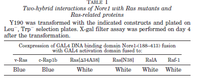
TABLE I Two-hybrid interactions of Nore1 with Ras mutants and Ras-related proteins

Detection of Ras/Nore1 Binding in Vitro—Purified, procaryotic recombinant c-Ha-Ras (2.5 mg/ml) was loaded with GTPgS (2 mM) or GDPbS (2 mM) at 37 °C for 15 min in the buffer containing 50 mM Tris-HCl, pH 7.5, 7.5 mM EDTA, 2.5 mM MgCl2 0.5 mg/ml bovine serum albumin, 1 mM dithiothreitol.

Various amounts of GTPgS- or GDPb Sloaded Ras proteins were mixed with purified procaryotic recombinant GST-Nore1-(188–413). Subsequent steps were essentially the same as described previously (12).

Detection of Ras-Nore1 Association in COS-7 Transient Expression System—COS-7 cells were plated at a density of 1.2 million/10-cm dish and transfected 24 h later with 7 mg of pMT2-HA-c-Ha-Ras or empty vector and 12 mg of pEBG-GST-Nore1 using the DEAE-dextran method.


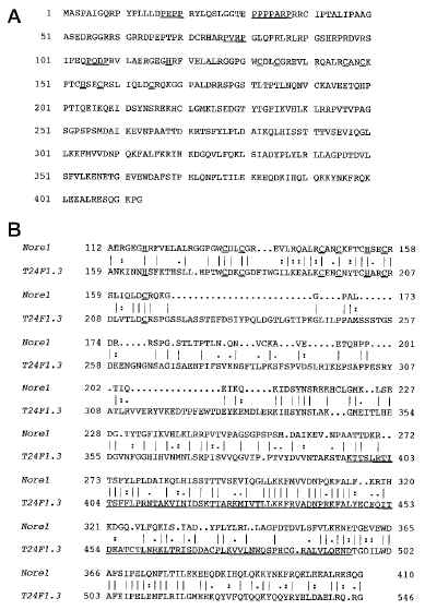
FIG. 1. Predicted protein sequence of Nore1 and alignment

FIG. 1. Predicted protein sequence of Nore1 and alignment with C. elegans gene product T24F1.3. A, the predicted protein sequence of Nore1. The open reading frame contains 413 amino acids. The DAG_PE binding domain and PXXP motifs are underlined. B, alignment of Nore1 with C. elegans gene product T24F1.3. The predicted RA (Ras/Rap association) domain (16) in T24F1.3 is underlined (aa 396–496). The GCG command BESTFIT was used to create the alignment. GAP creation and extension penalties were 4 and 2, respectively.

48 h later, cells were starved for 24 h and subsequently were stimulated with 100 ng/ml EGF for various times.

Cells were extracted in lysis buffer (30 mM HEPES, pH 7.4, 1% Triton X-100, 20 mM b-glycerophosphate, 2 mM NaPPi, 1 mM orthovanadate, 20 mM NaF, 20 mM KCl, 2 mM EGTA, 3 mM EDTA, 7.5 mM MgCl2, 14 mM b-mercaptoethanol, and a
mixture of protease inhibitors).

Lysates were freeze-thawed once and spun at 17,000 3 g for 20 min. Supernatants were incubated with anti-HA antibodies and protein A-G-Sepharose beads for 3–4 h at 4 °C and then washed extensively with lysis buffer. The washed beads were eluted in SDS sample buffer and the extracted proteins subjected to SDS-PAGE, transferred on PVDF membranes, and probed using the antibodies indicated.  Bound antibodies were visualized using ECL.

Detection of Ras-Nore1 Association in KB Cells—KB cells were grown to 80% confluence, starved of serum for 24 h, and subsequently stimulated with EGF (100 ng/ml) for various time. Lysates were prepared as for the COS-7 cells, and Ras was immunoprecipitated with anti-Ras antibody (Y13-238) and protein A-G-Sepharose beads. Subsequent steps were similar to those used in the COS-7 experiment.

Antibody Production—GST-Nore1-(188–413) was used to immunize New Zealand White rabbits. The antiserum was first depleted of GSTreacting antibodies by repeated incubation with immobilized GST. The GST-depleted antiserum was then affinity-purified using immobilized Nore1-(188–413) on a PVDF membrane.

RESULTS AND DISCUSSION
A yeast two-hybrid screen was carried out to identify potential new Ras effectors in mammalian cells. One million yeast
transformants coexpressing a V12 Ras bait plasmid and a cDNA library prepared from activated mouse T cells were screened. Twenty strong positives were obtained, which showed both interaction-dependent growth on selective media
and interaction-dependent expression of Lac-Z activity.

DNA sequencing revealed that 18 of the 20 positives were either mouse A-Raf or c-Raf-1. Two positive clones both encoded a 2.5-kb cDNA representing a new gene, which was named Nore1 (novel Ras effector). Although the sequence indicated that an incomplete open reading frame had been recovered, we made use of the yeast two-hybrid system to examine the specificity of the interaction of Nore1 with two Ras-related proteins Rap1b and RalA, and two well defined Ras effector domain mutants, Ras 12VD34,38A or Ras 12V38N, which are defective in binding known Ras effectors like Raf (12).

Rap1b and RalA belong to the Ras subfamily of small GTP-binding proteins (13); Rap1 (A and B) has identical sequence to Ras in the region corresponding to the Ras effector domain (aa 32–40) and binds to several previously identified Ras effectors like Raf, PI 3-kinase and Ral-GDS, whereas RalA does not bind to these polypeptides. Nore1 interacts with wild type Ras but not with Ras 12VD34,38A and Ras 12V38N. Nore1 also interact with Rap1b, but not with RalA (Table I).

Thus, the interactions of Nore1 with these Ras-related proteins parallels closely the pattern exhibited by other well established Ras effectors.

The 2.5-kb Nore1 cDNA insert was used as the hybridization probe to isolate the entire cDNA from a mouse brain cDNA
library. A 3018-base pair cDNA was isolated. The cDNA sequence around the first ATG matches the Kozak consensus
sequence for a translational start. The open reading frame from this methionine includes 413 amino acids, as shown in Fig. 1A, yielding a highly basic polypeptide (pI 5 9.41) with a predicted molecular mass of 46.4 kDa.

One obvious structural feature of Nore1 is the presence of a cysteine-histidine-rich segment typical of a diacylglycerol/phorbol ester (DAG_PE) binding site (14) (aa 118–165, H-X13-C-X2-C-X10-C-X2-C-X4-H-X2-C-X7-C).
Nore1 also has a proline-rich region in its amino-terminal region, with five PXXP sequences
(aa 17–20, PEPP; aa 31–34. PPPP; aa 34–37, PARP; aa 77–80, PVRP; and aa 105–108, PQDP),
which are possible SH3 domain binding sites (15).

A search of the GeneBank™ using BLASTP command found that a Caenorhabditis elegans gene product called T24F1.3 has significant homology to Nore1. Fig. 1B shows the sequence similarity; the most significant homology between the two proteins is in their carboxyl-terminal regions. T24F1.3 has been suggested previously to contain a Ras/Rap association domain (RA domain) (16), located at aa 396–496. This domain is within the region most closely related in sequence to Nore1.

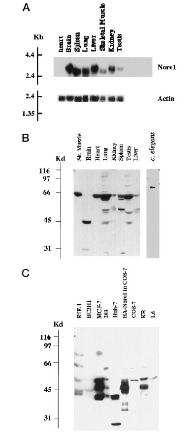
Nore1 mRNA abundance and complexity in murine tissues was examined by Northern blot (Fig. 2A). A single mRNA
generally about 3.1 kb was detected in most mouse tissues, although some size variation is noted. The highest levels are
observed in brain, liver, and spleen, with barely detectable levels in heart.

A polyclonal antibody was raised against a carboxyl-terminal fragment of Nore1 (aa 188–413) and purified by affinity chromatography using the recombinant antigen. Immunoblot of extracts prepared from different rat tissue is shown in Fig. 2B.


FIG. 2. Expression of Nore1 mRNA in mouse tissues and immunoreactive Nore1 polypeptides in rat tissues and cell lines.

FIG. 2. Expression of Nore1 mRNA in mouse tissues and immunoreactive Nore1 polypeptides in rat tissues and cell lines. A, expression of Nore1 mRNA in mouse tissues. A 220-base pair cDNA fragment (nucleotides 90–310) was labeled with [32P]dCTP by random priming method and used for probing Nore1 mRNA from various mouse tissues. The mouse multiple tissue blot was purchased from CLONTECH. B, protein expression of Nore1 in rat tissues. C, protein expression of Nore1 in cell lines. Cell lysates were all prepared in radioimmune precipitation buffer. Affinity-purified anti-Nore1 antibodies were used to probe the membrane. Bands were visualized by the ECL method..

A single immunoreactive band at 46 kDa is seen in a brain extract, which is in agreement with the predicted size of the polypeptide encoded by Nore1 cDNA isolated from the mouse brain library. A similar 46-kDa band is also seen in other tissues, including lung and testis. In addition, however, prominent immunoreactive bands at other molecular masses are seen in most tissues, and some tissues lack a 46-kDa band entirely (e.g. skeletal muscle, heart, spleen, and liver). All tissues but brain show a major 65-kDa band, and two bands around 55 kDa are also seen in lung, spleen, testis, and liver.

The 65- and 55-kDa bands may represent isoforms of Nore1, the existence of which is suggested by the partial cDNAs isolated from a variety of cDNA libraries (data not shown). Alternatively, these bands may reflect polypeptides unrelated to Nore1, except for the presence of sequence epitopes recognized by the polyclonal antibodies to Nore1.

The anti-Nore1 antibody also immunoblotted a single polypeptide in an extract prepared from C. elegans. This band is approximately 74 kDa, as compared with the molecular mass of T24F1.3 gene product of 69.1 kDa. The murine brain Nore1 cDNA was tagged at the Nore1
amino terminus with an HA epitope and expressed transiently in COS cells. As seen in Fig. 2C.

HA-Nore1 shows the expected size of 46 kDa by immunoblot with anti-Nore1 antibodies. Extracts prepared from several cell lines were subjected to Nore1 immunoblot; of the cell lines examined, only BC3H1, a vascular smooth muscle-like line derived from a radiationinduced murine brain tumor, shows a single band at 46 kDa.

 A band of similar size is seen in several other cell lines, including RIE-1 (rat intestinal epithelial), MCF-7 (human breast cancer), HEK 293 (human embryonic kidney), and KB (human oral carcinoma); however, immunoreactive polypeptides of 55 kDa (RIE-1, MCF-7, HEK 293, and KB) and 65 kDa (RIE-1, HEK 293, and KB), are as or more abundant in these cell lines, and some lines show only bands other than the 46-kDa polypeptide (e.g. Huh-7, 40 kDa; L6, 55 kDa).

We preabsorbed the affinitypurified anti-Nore1 antibodies with an excess amount of recombinant Nore1-(188–413) for 1 h and used this preabsorbed antibodies to probe the blots used in Fig. 2, B and C, and we did not see the predominant bands at 46, 55, and 66 kDa, suggesting that these bands in both figures are probably specific.

A GST-Nore1-(188–413) fusion protein (corresponding to the Nore1 polypeptide encoded in the initial cDNA isolate) was
expressed and purified from Escherichia coli. Procaryotic recombinant c-Ha-Ras was loaded with GTPgS or GDPbS, and
various amounts were mixed with a fixed amount of GSTNore1-( 188–413) or GST as control. After incubation at 30 °C
for 20 min, GST or GST fusion proteins and any associated proteins were recovered by addition of glutathione Sepharose beads. The beads were washed and eluted into SDS sample buffer; proteins were separated by SDS-PAGE, transferred to PVDF membrane, and probed for Ras using a monoclonal anti- Ras antibody.


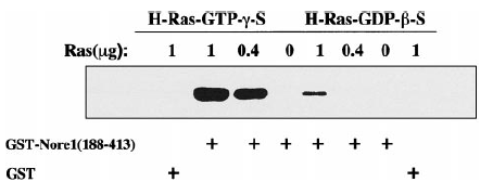
FIG. 3. Specific, GTP-dependent interaction of purified recombinant Nore1 and Ras polypeptides in vitro.

FIG. 3. Specific, GTP-dependent interaction of purified recombinant Nore1 and Ras polypeptides in vitro. V12-Ras protein purified from bacterial expression was loaded with either GTPgS or GDPbS. The loaded Ras proteins were incubated with GST or GSTNore1-(188–413). Glutathione-Sepharose beads were used to pull down the Ras-Nore1-(188–413) complexes. Ras protein was detected using the pan-Ras antibody-2 (Oncogene Science) in the Western blot shown above.

GST -Nore1-(188–413), but not GST binds Ras, and considerably more Ras-GTPgS is bound than Ras-GDP-b-S (Fig. 3). These results establish that the effector loop-dependent interaction between Nore1 and Ras identified by two-hybrid techniques reflects the direct binding of the two proteins and that the binding between Nore1 and Ras is GTP-dependent.

We then attempted to detect an interaction between Nore1 and Ras in mammalian cells and to determine whether this
binding was dependent on Ras activation in situ. 

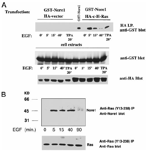
COS-7 cells were cotransfected with plasmids encoding GST-Nore1 and HA-tagged c-Ha-Ras. Forty-eight hours later, cells were serumstarved for 24 h and then stimulated with EGF or TPA for various times, extracted into buffer containing Triton X-100, and HA-Ras was recovered using the anti-HA monoclonal antibody, 12CA5. The washed immunoprecipitates were eluted into SDS sample buffer and separated by SDS-PAGE, transferred to PVDF membrane, and probed with affinity-purified anti-GST polyclonal antibodies. 

As seen in Fig. 4A, GST-Nore1 was specifically pulled down with HA-c-Ha-Ras, but only after the cells were treated with EGF or TPA; the expression of HA-c-Ha-Ras and of GST Nore1 was uniform throughout.

Thus, Nore1 is not detectably associated with Ras in serumstarved COS cells; however, within 5 min after stimulation by EGF (or TPA), Nore1 associates specifically with Ras; this association diminishes by 15 min after EGF addition and is largely reversed by 40 min, probably reflecting the down-regulation of Ras activation after EGF treatment.

We next attempted to detect an in situ association between endogenous Ras and endogenous Nore1, under conditions where the levels of the two polypeptides are not increased artificially by transient overexpression. We chose to examine the human oral carcinoma cell line KB, because Nore1 expression is readily detectable, and these cells express substantial numbers of EGF receptors. KB cells grown to 80% confluence were serum-starved for 24 h and then treated with EGF for various times.

Triton X-100-soluble cell lysates were subjected to immunoprecipitation using the monoclonal anti-Ras antibody, Y13-238, which are known to enable isolation of Ras-Raf complexes.

FIG. 4. EGF and TPA stimulated association of Nore1 with Ras in transfected COS-7 cells and in nontransfected KB cells.

FIG. 4. EGF and TPA stimulated association of Nore1 with Ras in transfected COS-7 cells and in nontransfected KB cells. A, EGF and TPA stimulated association of Nore1 with Ras in transfected COS-7 cells. pEBG-Nore1 together with PMT2-HA or PMT2-HA-c-Ha-
Ras were transiently expressed in COS-7 cells. Transfected cells were first serum-starved for 24 h and then stimulated with EGF (100 ng/ml) or TPA (100 nM) for the time indicated. Monoclonal anti-hemagglutinin antibodies were used to immunoprecipitate HA-c-Ha-Ras or HA alone. B, EGF stimulated association of endogenous Nore1 with endogenous Ras in KB cells. Confluent KB cells were serum-starved for 24 h and then stimulated with EGF (100 ng/ml) for various time. Triton X-100- soluble cell extracts were prepared, and the moloclonal anti-Ras antibody Y13-238 were used to immunoprecipitate Ras proteins.

The Ras immunoprecipitates were washed extensively with the lysis buffer, eluted into SDS sample buffer and subjected to SDS-PAGE, transferred to PVDF membrane, and immunobloted with the affinity-purified polyclonal anti-Nore1 antibodies.

As shown in Fig. 4B, although equal amounts of endogenous Ras were recovered in all samples, the Ras immunoprecipitates contain immunoreactive Nore1 only after treatment of the cells with EGF. The time course of Ras-Nore1 association after EGF treatment in KB cells is more sustained than that observed in COS-7 cells. This may reflect different time course of down-regulation of Ras activation in those cells. Interestingly, only the 46-kDa (and not the equally abundant 55-kDa) immunoreactive Nore1 polypeptide is recovered with c-Ras.

In summary, we have identified Nore1, a potential new Ras effector or target, using the yeast two-hybrid screen with Ras as bait. We show that Nore1 can bind Ras directly in vitro using purified recombinant Ras and Nore1 polypeptides. The Ras/ Nore1 association in vitro depends strongly on Ras being in the GTP-bound form. We show that with yeast two-hybrid assay, Nore1 interacts with Ras 12V but not two transformation defective effector loop mutants, Ras 12VD34,38A and Ras 12V38N.

This profile of interaction with Ras is identical to that exhibited by known and potential Ras effectors, including Raf, PI 3-kinase, Ral GDS, Rin1, and AF-6. We also show that the Ras/Nore1 association occurs in vivo following EGF and TPA activation of Ras in COS-7 cells overexpressing Ras and Nore1.

Finally, it is clear that a stimulus-dependent association of endogenous Ras and Nore1 occurs following EGF receptor activationin KB cells. To our knowledge, Nore1 is the only other candidate mammalian Ras effector, other than Raf, wherein the endogenous polypeptide has been demonstrated to associate with Ras in vivo following receptor activation. Taken together, these properties indicate that Nore1 is very likely to be a physiologic Ras effector.

* This work was supported in part by National Institutes of Health Grants GM51281 (to X.-F. Z.) and DAMD 17-94-54404 (to J. A.) and by a grant from Lilly Inc. (to J. A.) and ONASSIS (to D. V.). The costs of publication of this article were defrayed in part by the payment of page charges. This article must therefore be hereby marked “advertisement” in accordance with 18 U.S.C. Section 1734 solely to indicate this fact.

‡ To whom correspondence should be addressed. Tel.: 617-726-9450;
Fax: 617-726-9452; E-mail: zhang@helix.mgh.harvard.edu.

1 The abbreviations used are: EGF, epidermal growth factor; aa, amino acid(s); PI, phosphatidlyinositol; X-gal, 5-bromo-4-chloro-3-indolyl
b-D-galactopyranoside; kb, kilobase pair(s); GTPgS, guanosine 59-O- (thiotriphosphate); GDPbS, guanyl-59-yl thiophosphate; GST, glutathione S-transferase; PAGE, polyacrylamide gel electrophoresis; PVDF, polyvinylidene difluoride; HA, hemagglutinin; TPA, 12-O-tetradecanoylphorbol- 13-acetate; DAG, diacylglycerol.

References

1. Fantl, W. J., Johnson, D. E., and Williams, L. T. (1993) Annu. Rev. Biochem.
62, 453–481
2. Woodrow, M. A., Rayter, S., Downward, J., and Cantrell, D. A. (1993)
J. Immunol. 150, 3853–3861
3. Van Corven, E. J., Hordijk, P. L., Medema, R. H., Bos, J. L., and Moolenaar,
W. H. (1993) Proc. Natl. Acad. Sci. U. S. A. 90, 1257–1261
4. Wittinghofer, A., and Nassar, N. (1996) Trends Biochem. Sci. 21, 488–491
5. Avruch, J., Zhang, X.-F., and Kyriakis, J. M. (1994) Trends Biochem. Sci. 19,
279–283
6. Cowley, S., Paterson, H. Kemp, P., and Marshall, C. J. (1994) Cell 77, 841–852
7. Oldham, S. M., Clark, G. J., Gangarosa, L. M., Coffey, R. J. Jr., and Der, C. J.
(1996) Proc. Natl. Acad. Sci. U. S. A. 93, 6924–6928
8. D’Arcangelo, G., and Halegoua, S. (1993) Mol. Cell. Biol. 13, 3146–3155
9. Taylor, S. J., and Shalloway, D. (1996) Curr. Biol. 6, 1621–1627
10. Katz, M. E., and McCormick, F. (1997) Curr. Opin. Genet. Dev. 7, 75–79
11. Marshall, C. J. (1996) Curr. Opin. Cell Biol. 8, 197–204
12. Zhang, X.-F., Settleman, J., Kyriakis, J. M., Takeuchi-Suzuki, E., Elledge,
S. J., Marshall, M. S., Bruder, J. T., Rapp, U. R., and Avruch, J. (1993)
Nature 364, 308–313
13. Valencia, A., Chardin, P., Wittinghofer, A., and Sander, C. (1991) Biochemistry
30, 4637–4648
14. Ono, Y., Fujii, T., Igarashi, K., Kuno, T., Tanaka, C., Kikkawa, U., and
Nishizuka, Y. (1989) Proc. Natl. Acad. Sci. U. S. A. 86, 4868–4871
15. Ren, R., Mayer, B. J., Cicchetti, P., and Baltimore, D. (1993) Science 259,
1157–1161
16. Ponting, C. P., and Benjamin, D. R. (1996) Trends Biochem. Sci. 21, 422–425

Identification of Nore1 as a Potential Ras Effector
Demetrios Vavvas, Xin Li, Joseph Avruch and Xian-Feng Zhang

J. Biol. Chem. 1998, 273:5439-5442.
doi: 10.1074/jbc.273.10.5439

Access the most updated version of this article at http://www.jbc.org/content/273/10/5439
Alerts:
• When a correction for this article is posted
• When this article is cited
Click here to choose from all of JBC’s e-mail alerts
http://www.jbc.org/content/273/10/5439.full.html#ref-list-1
This article cites 16 references, 6 of which can be accessed free at
http://www.jbc.org/content/273/10/5439.full.html#ref-list-1

Diagnosis ⁄ Therapy in Ophthalmology

Diagnosis ⁄ Therapy in Ophthalmology

Demetrios G. Vavvas, Nancy Huynh,
Louis Pasquale and Eliot L. Berson.

Massachusetts Eye and Ear Infirmary.
Department of Ophthalmology
Harvard Medical School | Boston, Massachusetts, USA

Acta Ophthalmol. 2010: 88: 156–157
2008 The Authors Journal compilation
ª 2008 Acta Ophthalmol
doi: 10.1111/j.1755-3768.2008.01359.x

A70-year-old woman presented with a complaint of diminishing vision over 2 years, which was worse in the left eye (OS). She had received hydroxychloroquine treatment for discoid lupus for approximately 15 years at doses of < 400 mg⁄ day (< 6.5 mg⁄ kg ⁄ day; cumulative dose of  ≈ 2.2 kg).

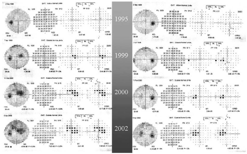
The medication was discontinued in 2000 at the onset of ocular symptoms.  Serial visual fields (VFs) (Fig. 1) revealed progressive loss that had prompted one ophthalmologist to initiate treatment for normal-tension glaucoma. 

Fig. 1. Humphrey visual field (VF) findings in a 70-year-old woman with hydroxychloroquine retinal toxicity showing progressive VF loss from 1999–2002. The patient stopped hydroxychloroquine in 2000.

Fig. 1. Humphrey visual field (VF) findings in a 70-year-old woman with hydroxychloroquine retinal toxicity showing progressive VF loss from 1999–2002. The patient stopped hydroxychloroquine in 2000.

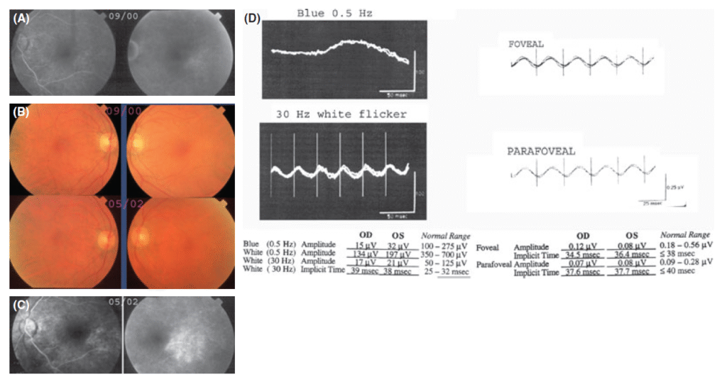
Fluorescein angiography (FA) in 2000 (Fig. 2A) showed areas of hyperfluorescence in the parafoveal region, leading to an alternative diagnosis of juxtafoveal telangiectasia.

Fig. 2. A Fluorescein angiogram FA in 2000 OS with early and late frames showing hyperfluorescence in the parafoveal area. B Fundus

Fig. 2. (A) Fluorescein angiogram (FA) in 2000 (OS) with early and late frames showing hyperfluorescence in the parafoveal area. (B) Fundus

On examination, the patient’s visual acuity was 20 ⁄ 20 OD and 20 ⁄ 60 OS. No afferent pupillary defect was seen. Intraocular pressures
were 14 mmHg OD and 15 mmHg OS. No colour deficit on Ishihara plates was found. Examinations of anterior segments, optic discs and
maculae were normal (Fig. 2B).  Repeat VF testing and FA findings were similar to those obtained previously (Fig. 2A, C). Scotopic and
photopic electroretinography (ERG) revealed suppressed and delayed photoreceptor responses, suggestive of widespread outer retinal disease that is unexpected in glaucoma (Fig. 2D). Hydroxychloroquine is an antimalarial drug frequently used in the treatment of haematological diseases.

One of its potentially adverse sideeffects is irreversible maculopathy, although this is rare relative to the number of patients on the drug (Levy et al. 1997).  Early signs of hydroxychloroquine toxicity can present as a paracentral scotoma detected by threshold VF testing. Advanced toxicity is typically characterized by bull’s eye maculopathy associated with retinal pigment epithelium (RPE) atrophy (Easterbrook 1999).

Our patient developed hydroxychloroquine toxicity over 2 years after discontinuation of the medication, with incomplete bull’s eye
lesions only apparent on FA. The pattern of VF loss mimicked an arcuate defect seen in glaucoma. However, it did not respect the horizontal meridian despite adequate patient fixation, as shown by the position of the blind spot (Fig. 1).

Finally, glaucoma, as a disease that involves the ganglion cells, does not affect full-field or focal ERG amplitude responses to single flashes of light. Our patient’s ERG was diminished, which is in accordance with hydroxychloroquine toxicity (Weiner et al. 1991).

The FA findings, showing RPE changes and the absence of telangiectatic vessels or leakage, were also consistent with the diagnosis of hydroxychloroquine retinopathy, but not with juxtafoveal telangiectasis.

References

Easterbrook M (1999): Detection and prevention of maculopathy associated with antimalarial agents.
Int Ophthalmol Clin 39: 49–57.
Levy GD, Munz SJ, Paschal J, Cohen HB, Pince KJ & Peterson T (1997): Incidence of hydroxychloroquine retinopathy in 1207 patients in a large multicentre outpatient practice. Arthritis Rheum 40: 1482–1486.
Weiner A, Sandberg MA, Gaudio AR, Kini MM & Berson EL (1991): Hydroxychloroquine retinopathy. Am J Ophthalmol 112: 528–534.

Regression of Some High-risk Features of Age-related Macular Degeneration (AMD) in Patients Receiving Intensive Statin Treatment

Regression of Some High-risk Features of Age-related Macular Degeneration (AMD) in Patients Receiving Intensive Statin Treatment

Research Paper

Regression of Some High-risk Features of Age-related Macular Degeneration (AMD) in Patients Receiving Intensive Statin Treatment

Demetrios G. Vavvas ⁎  Anthony B. Daniels ⁎ Zoi G. Kapsala, JeremyW. Goldfarb, Emmanuel Ganotakis, John I. Loewenstein, Lucy H. Young, Evangelos S. Gragoudas, Dean Eliott, Ivana K. Kim Miltiadis K. Tsilimbaris ⁎ JoanW. Miller ⁎

Retina Service, Department of Ophthalmology, Mass. Eye and Ear Infirmary, Harvard Medical School, Boston, MA, USA
Retina Service, Department of Ophthalmology, University of Crete, Heraklion, Crete, Greece

1. Introduction
Age-related macular degeneration (AMD) is the leading cause of irreversible vision loss in the developed world (Miller, 2013; Wong
et al., 2014). The non-neovascular or “dry” form accounts for 85% of all AMD and is characterized by accumulation of extracellular deposits, termed drusen (Sarks et al., 1994), between the basal lamina of retinal pigmented epithelium (RPE) and inner collagenous layer of Bruch’s membrane (BM), which is the inner wall of the choroid. Progression to advanced AMD involves atrophy of the RPE and overlying photoreceptors (geographic atrophy), and/or choroidal neovascularization (neovascular or “wet” AMD).While there are effective anti-angiogenic therapies for the less prevalent neovascular AMD, there are no effective treatments for the more prevalent dry form (Miller, 2013).